
Extended Data Figure 10. Presence and absence of collaterals from NOT-DTN-projecting cortical neurons in selected brain areas.
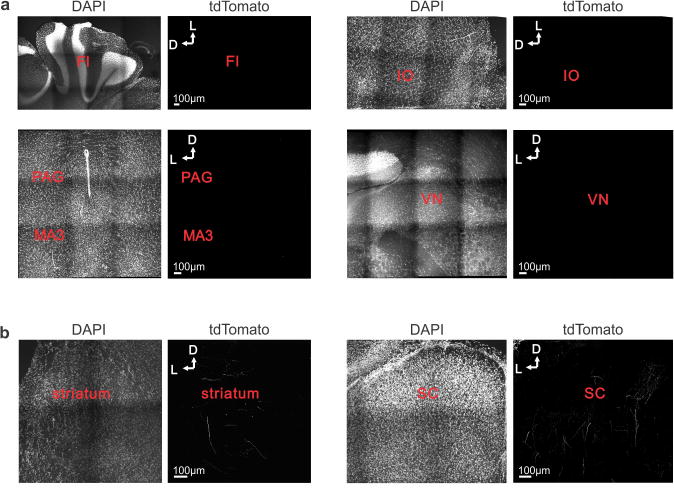
a, Absence of collaterals from NOT-DTN-projecting cortical neurons in flocculus (FL); inferior olive (IO); periaqueductal grey (PAG); medial accessory oculomotor nucleus (MA3); and vestibular nuclei (VN). For each coronal section the left panel is the DAPI fluorescence signal (blue channel) and the right panel is the tdTomato fluorescence signal (red channel). b, Presence of collaterals from NOT-DTN-projecting cortical neurons in the striatum and superior colliculus (SC).