
Figure 2. Cortico-fugal projection from mouse visual cortex to NOT-DTN.
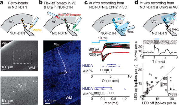
a–d, Top, schematic of experimental design. VC, visual cortex. a, Beads injected into NOT-DTN are retrogradely transported to visual cortex. Middle, coronal slice of visual cortex. Bottom, higher magnification of the enclosed area. WM, white matter. b, Example of layer 5 pyramidal cell projecting to NOT-DTN in visual cortex. Blue, DAPI; white, tdTomato. c, Middle, AMPA receptor-mediated (downwards) or NMDA receptor-mediated (upwards) EPSCs evoked in NOT-DTN neurons by optogenetic stimulation of cortico-fugal axons in vitro. Black, individual traces; red, average traces; blue, time course of blue light illumination. Bottom, summary of onset latency and trial-by-trial jitter of EPSCs. Data shown as mean ± s.d.; n = 9 mice. d, Firing of NOT-DTN neurons upon optogenetic activation of visual cortex in vivo. Middle, raster plot and PSTH of a NOT-DTN unit. Blue bar, LED illumination. Bottom, firing rates of NOT-DTN units in LED-off trials vs LED-on trials. Dotted line, unity line. Inset, summary of onset latency (n = 23 units). Data shown as median ± s.d.; n = 4 mice.