
Figure 3. Cortico-fugal projection to NOT-DTN is necessary for OKR potentiation.
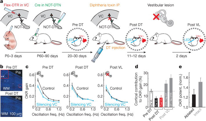
a, Schematic of experimental design. DTR, diphtheria toxin receptor; DT, diphtheria toxin; VL, vestibular lesion. b, Coronal slices of visual cortex from a mouse without diphtheria toxin injection (top) or with diphtheria toxin injection (bottom). Inset, higher magnification of the enclosed area. Blue, DAPI; white, GFP. c, Data from example mouse. Oscillation frequency tuning curves measured before ablation of the cortico-fugal projections (Pre DT, n = 60 trials), after ablation (Post DT, n = 30 trials) and after vestibular lesion (Post VL, n = 42 trials). d, Population average of cortical contribution to OKR gain. White, intact cortico-fugal projection. Black, after ablation of the cortico-fugal projection and before vestibular lesion. Red, after vestibular lesion. Same set of animals (n = 18 mice). Grey columns, two control groups after vestibular lesion: left, without diphtheria toxin injection (n = 6 mice); right, without DTR infection (n = 8 mice). e, Population average of OKR potentiation for mice with intact or ablated cortico-fugal projections (n = 27 and 18 mice, respectively). Data in c–e shown as mean ± s.e.m.