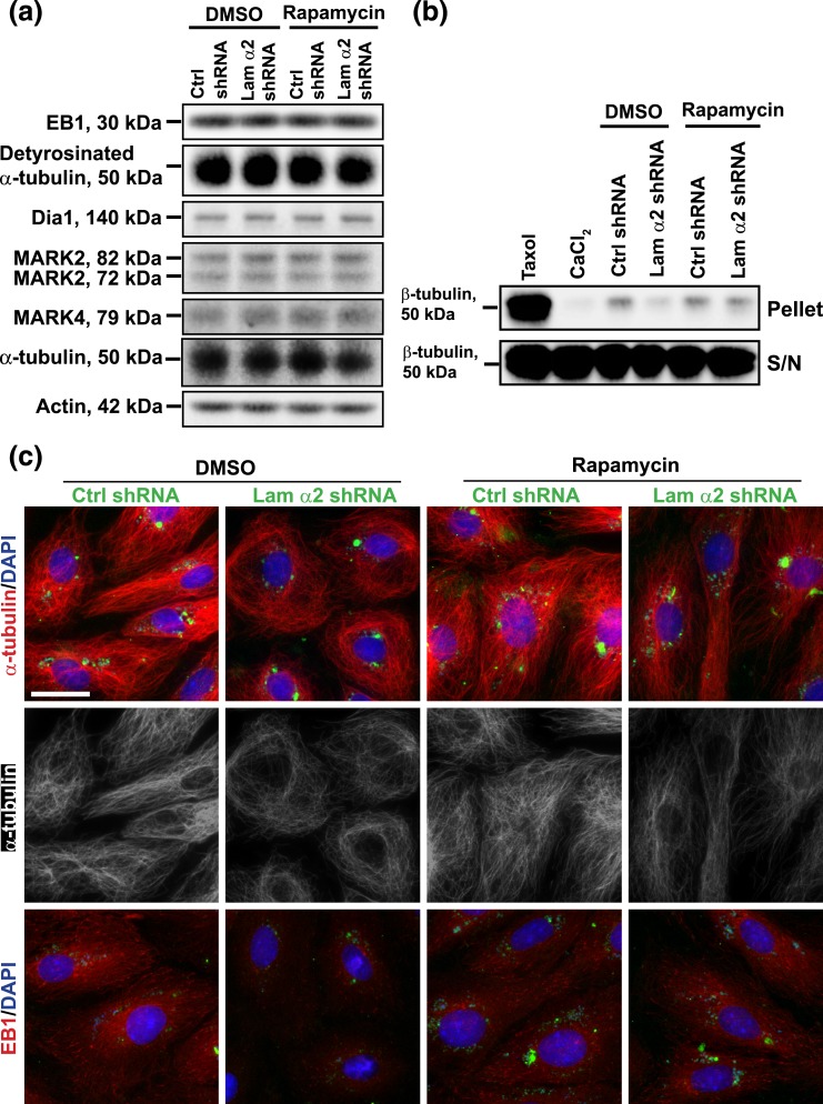
Figure 5.
Laminin α2 knockdown by shRNA that causes Sertoli cell MT disorganization is mediated through mTORC1 signaling. Sertoli cells cultured for 3 days were transfected with laminin α2 (Lam α2) vs negative control (Ctrl) shRNA for 24 hours. Cells were rinsed and treated with rapamycin (100 ng/mL) for 24 hours before harvested for IF analysis. For IB and MT polymerization assay, transfected cells were cultured for an additional 24 hours and were then treated with rapamycin (100 ng/mL) for 24 hours. (a) Studies by IB illustrated that laminin α2 knockdown without or with rapamycin treatment had no apparent effect on the expression of MT regulatory proteins EB1, MARK2 and MARK4 vs detyrosinated α-tubulin (the stabilized form of MTs, rendering MTs less dynamic). (b) MT polymerization assay was performed to quantify the ability of cell lysates to induce MT polymerization in Sertoli cells. Laminin α2 knockdown reduced MT polymerization because considerably less polymerized MTs were detected. However, treatment of Sertoli cells with rapamycin that blocked the function of mTORC1 abolished the laminin α2 knockdown-induced downregulation of MT polymerization (see pellet). The supernatant contained the monomers of MTs which were similar in all groups. Taxol (paclitaxel) and CaCl2 served as the corresponding positive and negative controls, which promoted and inhibited MT polymerization, respectively. (c) Studies by IF showed that laminin α2 knockdown led to MT disorganization as well as mislocalization of EB1 [a +TIP (plus-end tracking protein)] in which EB1 no longer prominently localized with MTs appeared to discrete dots along the MTs. In control cells, α-tubulin, building blocks of MTs, stretched across the entire cell cytosol, whereas MTs retracted from cell cytosol but rounded up to enclose the Sertoli cell nuclei after laminin α2 knockdown. On the other hand, EB1 associated with long stretches of MTs in control cells, but it no longer prominently found to associate with MTs after laminin α2 knockdown, likely dispersed in cell cytosol because the EB1 level was not perturbed based on IB [see (a)]. Rapamycin treatment that blocked the mTORC1 signaling rescued laminin α2 knockdown induced MT disorganization. GFP expression (green) illustrated the successful transfection. Sertoli cell nuclei were visualized by DAPI. Scale bar, 30 μm.

