Figure 3.
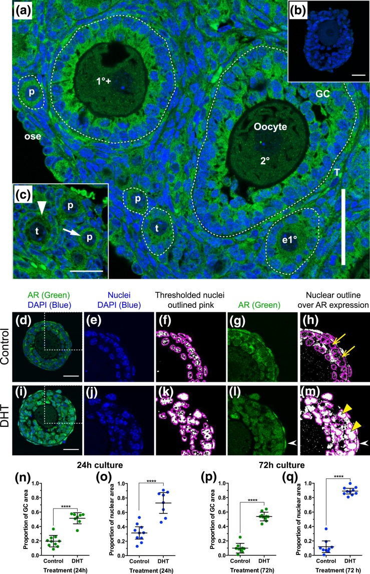
AR is present in oocytes, GCs, and theca cells of preantral follicles, and overall expression and nuclear localization of AR increases in the presence of DHT. (a) AR expression (green) in mouse prepubertal mouse ovary on day 17 postpartum. DAPI-labeled nuclei are blue. Scale bar = 50 µm. White dotted lines indicate basal lamina surrounding follicles. (b) Rabbit immunoglobulin G control. Scale bar = 25 µm. (c) Strong labeling of ooplasm (white arrow) and GC cytoplasm (white arrowhead) in primordial and transitional follicles. Scale bar is 25 µm. (d–m) Quantification of AR in formalin-fixed, paraffin-embedded sections of follicles cultured in the absence (d–h) and presence (i–m) of DHT. (d and i) Confocal image of follicle showing expression of AR (green) and DAPI-labeled nuclei (blue). Scale bars = 25 µm. White dotted box defines enlarged images (e–h and j–m). Images were imported into ImageJ and split into red-green-blue channels. Blue DAPI-labeled nuclei (e and j) were thresholded (f and k; where white is DAPI positive and black DAPI negative), and nuclei were outlined using the “Analyze Particles” command (f and k). In the green channel, AR labeling (g and l) was thresholded (with the same threshold maintained for all follicles), the nuclear outlines were overlaid, and the area of labeling exceeding the threshold was measured within the nuclear outlines (h and m), and for the GC layer as a whole. Yellow arrows (h) indicate nuclei with little nuclear AR, and yellow arrowheads (m) indicate nuclei with strong nuclear AR. White arrowheads (l and m) show strong AR expression in theca cell nuclei. Values were plotted and compared between treatments using a Mann-Whitney test. Overall AR protein expression in the GC layer was significantly increased after 24 hours (n) and 72 hours (p) culture in the presence of DHT. Nuclear localization of AR was also significantly increased in the presence of DHT at 24 hours (o) and 72 hours (q). 1°+, primary stage with second layer of GCs appearing; 2°, secondary stage; e1°, early primary; ose, ovarian surface epithelium; p, primordial; T, theca; t, transitional.

