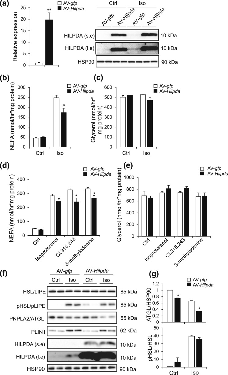
Figure 8.
Overexpression of Hilpda in adipocytes suppresses NEFA release. (a) Hilpda mRNA (left) and HILPDA protein levels (right) in fully differentiated 3T3-L1 adipocytes transduced with AV-gfp or AV-Hilpda. Fully differentiated 3T3-L1 adipocytes were trypsinized, replated at 70% confluency, serum starved for 24 hours, and transduced with recombinant AVs expressing gfp or Hilpda at a multiplicity of infection of 750 for 72 hours. Gene expression levels of AV-gfp–treated adipocytes were set at one. (b) NEFA and (c) glycerol release in fully differentiated 3T3-L1 adipocytes that were trypsinized, replated at 70% confluency, serum starved for 24 hours, and transduced with recombinant AVs expressing gfp or Hilpda at a multiplicity of infection of 750 for 72 hours. Transduced differentiated 3T3-L1 adipocytes were serum starved for 2 hours and incubated with 5 µM isoproterenol for 3 hours. (d) NEFA and (e) glycerol release in fully differentiated 3T3-L1 adipocytes that were transduced with recombinant AVs expressing gfp or Hilpda and incubated either with 10 µM isoproterenol, 5 mM 3-methyladenine, or 10 µM CL316,243 for 3 hours. (f) Representative immunoblots for HSL (LIPE), phospho-HSL, ATGL (PNPLA2), perilipin 1 (PLIN1), and HILPDA in fully differentiated 3T3-L1 adipocytes that were transduced with recombinant AVs expressing gfp or Hilpda and incubated with 10 µM isoproterenol for 3 hours. (g) Quantification of phospho-HSL and ATGL immunoblots in differentiated 3T3-L1 adipocytes, transduced as described earlier. Data are mean ± standard error of the mean. Asterisks indicate significant differences according to Student t test relative to AV-gfp–treated adipocytes; **P < 0.01; *P < 0.05. Ctrl, control; Iso, isoproterenol; l.e., long exposure; s.e., short exposure.

