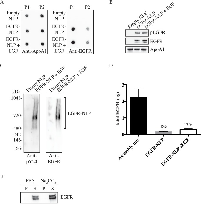
Fig 3. EGFR incorporation in self-assembled NLPs.
A, P1 and P2 SEC fractions were collected and spotted onto nitrocellulose membrane followed by immunoblotting with anti-ApoA1 and anti-EGFR antibodies showing that the majority of EGFR is incorporated into the higher molecular weight NLPs of the P1 fraction. B, Indicated NLPs were separated by denaturing SDS-PAGE and analyzed with anti-pEGFR, anti-EGFR, and anti-ApoA1 antibodies. Numbers indicate band intensity relative to EGFR-NLP normalized to ApoA1 signal. C, Purified Empty and EGFR-NLPs were separated by NativePAGE gels and immunostained with anti-pY20 and anti-EGFR antibodies showing EGFR expression in the higher molecular weight ranges above 480 kDa. D, Bar graph of determination of EGFR insertion into NLP. Total amount of EGFR in the NLP assembly mixture and purified EGFR-NLPs were determined by ELISA and EGFR insertion rate indicated. The values represent the mean ± standard error of the mean (SEM) for two technical replicates. E, EGFR-NLPs were extracted with PBS (control) or sodium carbonate (Na2CO3), centrifuged to separate the supernatant containing NLPs (S) and pellet containing insoluble free protein (P), separated by denaturing SDS-PAGE, and analyzed with anti-EGFR antibody showing that EGFR is not susceptible to carbonate extraction.

