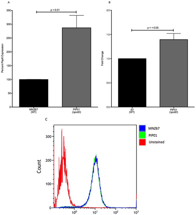
Fig 2. PspK analysis.
PspK was quantified in pneumococcal lysates by an indirect ELISA, and pspK expression was determined via RT-qPCR. Surface-expressed PspK was assessed by flow cytometry. Significantly more PspK was produced in potD mutant PIP01 when compared to wild type MNZ67 (A). Additionally, significantly higher pspK expression was seen in PIP01 (B). Although increased PspK was observed, no difference was seen in surface associated PspK (C). The ELISA was performed at least three times, and each sample was analyzed in triplicate. RT-qPCR was performed three independent times and data was analyzed using the ΔΔCT method. Flow cytometry was performed twice. Error bars denote standard error of the mean.

