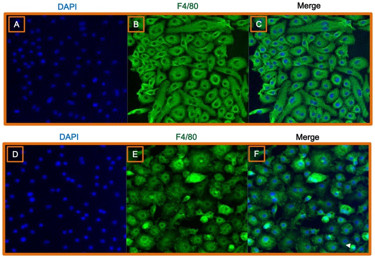
Fig 4. Immunofluorescent characterization of hepatic macrophages following C. sinensis infection.
Fluorescence images of hepatic macrophages harvested from uninfected (A–C) and C. sinensis-infected mice (D–F). Cells were stained with DAPI (blue; A, D) or an F4/80-specific antibody (green; B, E). Cells were observed by confocal microscopy at a magnification of 200×. (C, F) Merged images.

