
Figure 3.
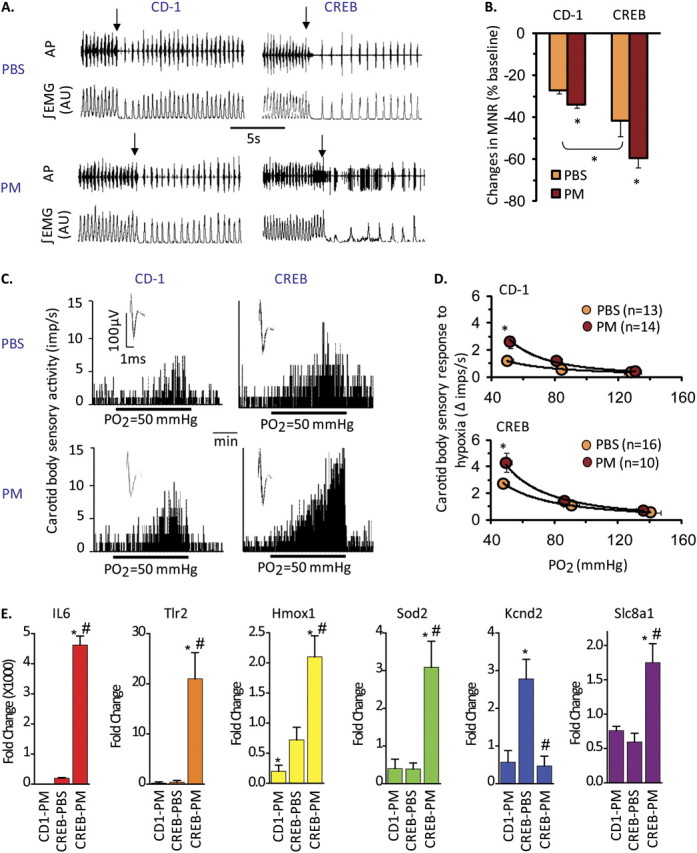
PM sensitizes carotid body function in CREBA133 mice. (A) Diaphragmatic EMG responses to brief (20 s) exposure to 100% O2 (arrows) in CREBA133 and CD1 mice exposed to vehicle (PBS) or PM. AP = action potential of the diaphragmatic EMG; AU = arbitrary units; ∫EMG = integrated diaphragmatic EMG activity. (B) Effects of brief hyperoxia (Dejour's test) on minute neural respiration (MNR). Data are mean ± SEM change in MNR (MNR100% –MNR21% O2)/MNR21%O2. n = 4 mice per group (*P < 0.05). (C and D) Integrated sensory activities of the carotid body in response to hypoxia in CD1 and CREBA133 mice exposed to PBS or PM (Po2 = partial pressures of O2 in the perfusate [Po2 = 50 mm Hg]). Black bars represent duration of the hypoxic challenge. The superimposed action potential of a “single” fiber is shown (inset). Average sensory responses to graded hypoxia are presented as changes in impulses/second (hypoxia – baseline). Values are mean ± SEM. *P < 0.05. n = Number of fibers analyzed from five mice per group. (E) PM induces dysregulation of inflammatory, oxidant, and carotid body regulatory genes in CD1 and CREBA133 mice. Pooled carotid body RNA from PBS- and PM-challenged CD1 and CREBA133 mice (36 h; n = 10–12 mice per group) was used in qPCR reactions (see Materials and Methods). Shown are expression levels for IL-6, Toll-like Receptor 2 (TLR2), heme oxygenase (Hmox1), superoxide dismutase (SOD2), the large conductance Ca2+–activated K+ channel (Kcnd2), and the Na+–Ca2+ exchanger 1 precursor protein (Slc8a1). Data represent the fold change in gene expression compared with PBS-challenged CD1 mice (*P < 0.05).