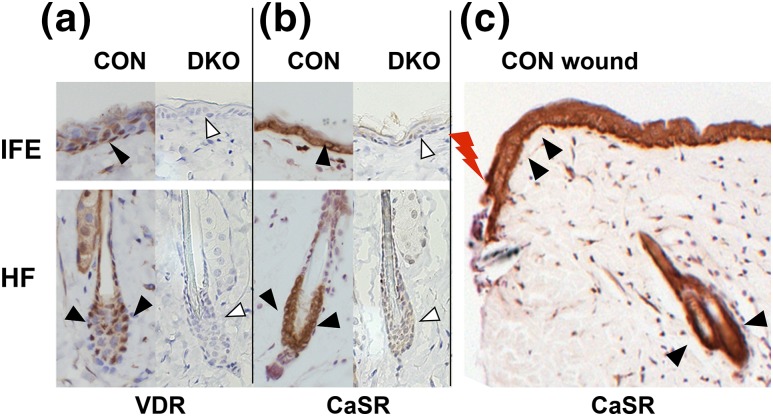
Figure 1.
DKO mice were generated in which both Vdr and Casr were removed from Krt14-expressing keratinocytes. Immunohistochemistry for (a) Vdr and (b, c) Casr in 3-month-old DKO skin (arrowheads) and littermate CON skin. The Casr staining in the epidermis at the edge of the wound (lightning bolt) in CON skin is also shown. Representative images of staining in IFE and HF are shown.

