Figure 5.
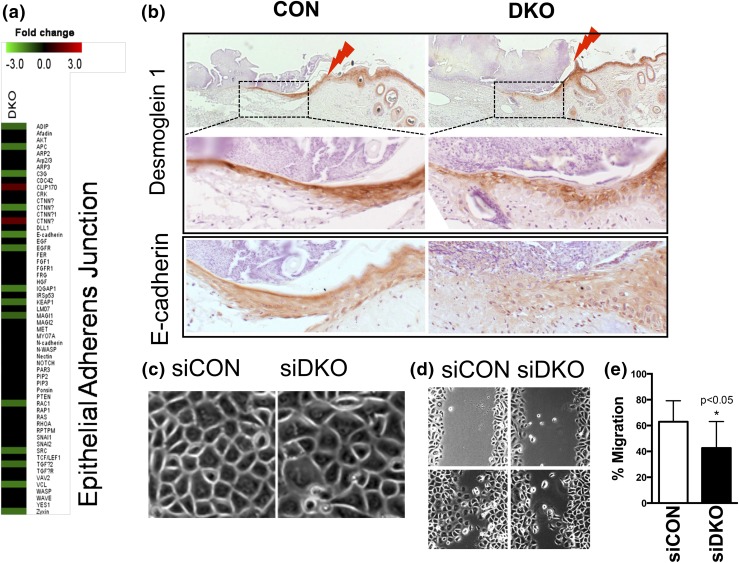
Deletion of both Vdr and Casr (DKO) impairs adhesion and migration of keratinocytes to re-epithelize the wound. (a) DKO downregulation of AJ signaling in epidermis as shown in microarray analyses. (b) Immunohistochemistry for desmoglein 1 (upper two panels) and E-cadherin (bottom panels) on the wound sections of CON (left) and DKO (right) mice (3 days after wounding at 3 months of age). The upper panels for desmoglein 1 are of low magnification, with the boxed regions shown in the middle panels at higher magnification. The lightning bolts show wound margins. Representative images from two analyses are shown. (c) The phase-contrast morphology of human keratinocytes transfected by siDKO compared with siCON (phase microscopy). (d, e) Migration capability was assessed by the scratch assay. Confluent cultures of transfected cells were treated to block proliferation, and they were scratched by pipette tips. After 16 hours, the migration rate was quantitated by measuring the amount of open area remaining in the scratch region through Bioquant software, and the results expressed as the ratio of closure after 16 hours to the original area of the scratch. Mean ± SD shown (n = 12). *P < 0.05.

