Figure 2.
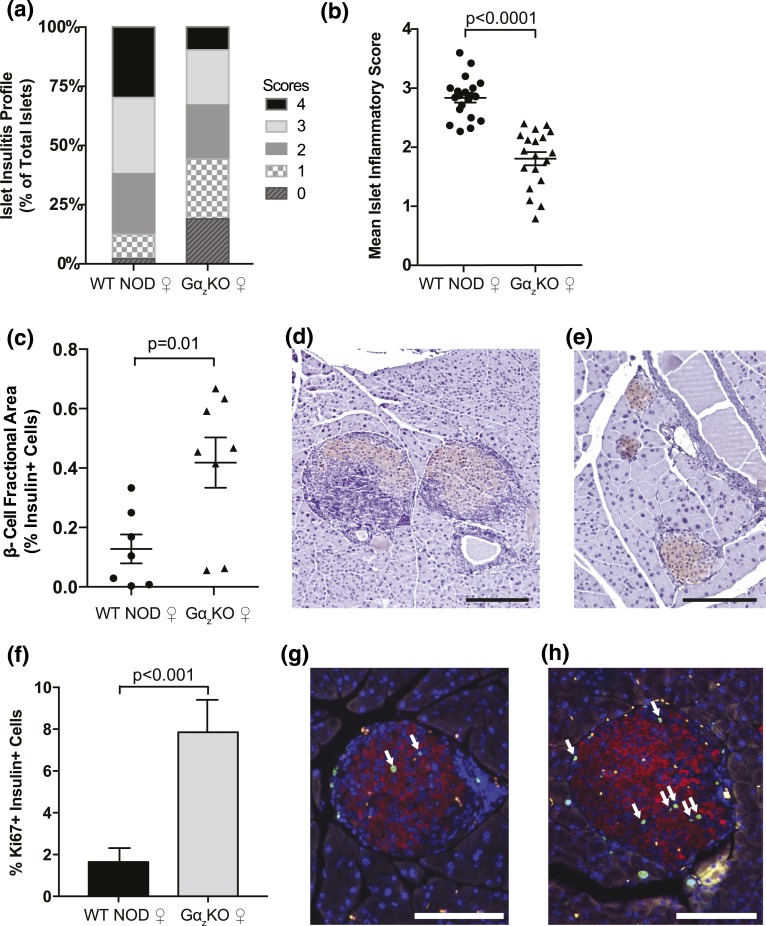
Female (♀) Gαz-KO NOD mice have decreased islet insulitis and increased insulin-positive pancreas area at 17 weeks of age. Pancreata were collected from female WT and Gαz-KO mice at 17 weeks of age and subjected to hematoxylin and eosin staining to determine islet inflammation or insulin immunohistochemistry with hematoxylin counterstain to determine β-cell fractional area. (a) Percentage of islets in each of the islet inflammation scoring categories. (b) Mean islet immune infiltration score for each biological replicate. (c) Quantification of β-cell fractional area calculated from all islets counted on three sections of each mouse pancreas separated by at least 200 μm. Representative pancreas sections from WT and Gαz-KO mice are shown in panels (d) and (e), respectively. (f) Quantification of insulin+ and Ki67+ cells in 17-week-old female mice calculated by counting all islets in a pancreas section and averaging two sections separated by at least 200 μm per mouse. Representative pancreas sections from WT and Gαz-KO mice are shown in panels (g) and (h), respectively. White arrows indicate insulin+ Ki67+ cells. In all images, the scale bar represents 200 μm. (b) Data were compared by Student t test; n = 21 per group. (c) n = 7 per group. (f) n = 6 per group.

