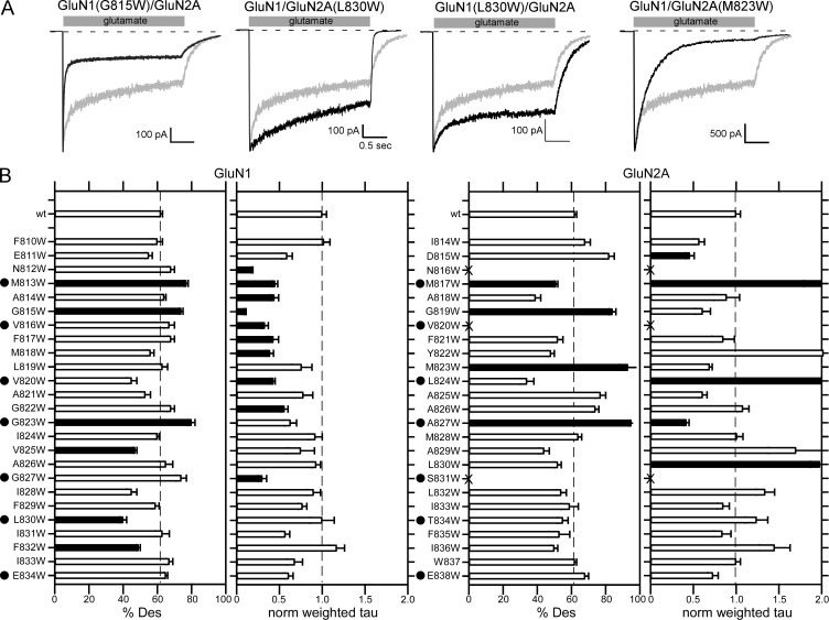
Figure 7.
Impact of M4 substitutions on NMDAR desensitization. (A) Representative whole-cell recordings of current through wild-type GluN1/GluN2A (gray traces) or NMDARs containing a tryptophan substitution in the M4 segment (black traces). Currents were recorded as in Fig. 3. (B) Mean (±SEM) of the %Des or normalized weighted τ for wild-type GluN1/GluN2A or GluN1/GluN2A containing tryptophan substitutions in either the GluN1 (left) or GluN2A (right) M4 segments. Raw values and details of number of recordings made are shown in Table 5. Solid bars indicate values significantly different from wild type (P < 0.01, t test). We used a more stringent level of significance for this analysis to focus on only those positions with prominent effects on desensitization. Dots highlight positions homologous to the VVLGAVE face in AMPARs.

