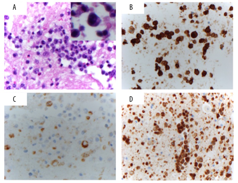
Figure 2.
Histology and Immunohistochemical (IHC) studies. (A) Cell block stained with hematoxylin and eosin (40×) showing atypical lymphocytes with enlarged nuclei and prominent nucleoli (inset, 100× magnification of an atypical large cell). (B) CD30 stains the majority of the cells (40×). (C) EMA stains a subset of cells, mainly the most atypical enlarged cells (40×). (D) CD3 stains most of the cells (40×).

