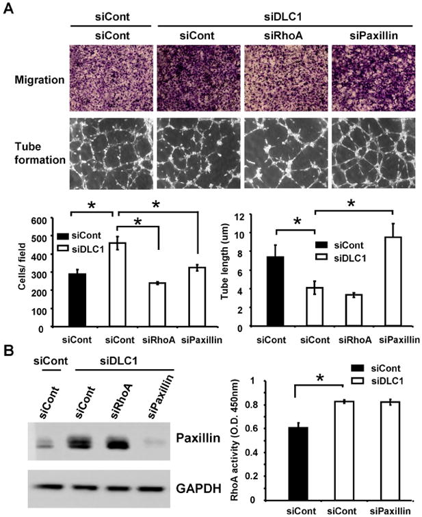
Figure 2. RhoA and paxillin independently contribute to the cell migration phenotype but only paxillin is involved in tube formation defects in siDLC1 HUVECs.

(A) Representative images of transwell cell migration (upper) or tube formation (lower) of HUVECs treated with indicated siRNAs and bar graphs of data from triplicate experiments. *, P < 0.05 (B) Cell lysates isolated from HUVECs treated with indicated siRNAs were immunoblotted with paxillin or GAPDH antibodies (left) or were used for measuring RhoA activities by Rho G-LISA activation assay (right)(N=3).
