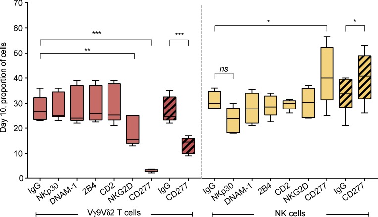
Figure 5.
A role for CD277 and NKG2D in the activation of Vγ9Vδ2 T cells by type I EBV infection. PBMCs from eight group 1 donors were preincubated with antagonistic mAbs against NKp30, DNAM-1, 2B4, CD2, NKG2D, CD277, or a control mouse IgG. They were then stimulated with Akata for 10 d in the presence of IL2. In other cultures, PBMCs alone were stimulated for 10 d with Akata cells preincubated with anti-CD277 or IgG control. Boxes and whiskers represent the proportion of Vγ9Vδ2 T cells (orange) and NK cells (yellow) after the 10-d culture with Akata cells. Hatched boxes and whiskers represent the proportion of Vγ9Vδ2 T cells (orange) and NK cells (yellow) after the 10-d culture with Akata cells preincubated with anti-CD277 or IgG control. Statistical significance of the difference between experiment and control was assessed using ANOVA (*, P = 0.01; **, P < 0.003; ***, P < 0.0001; ns, nonsignificant).

