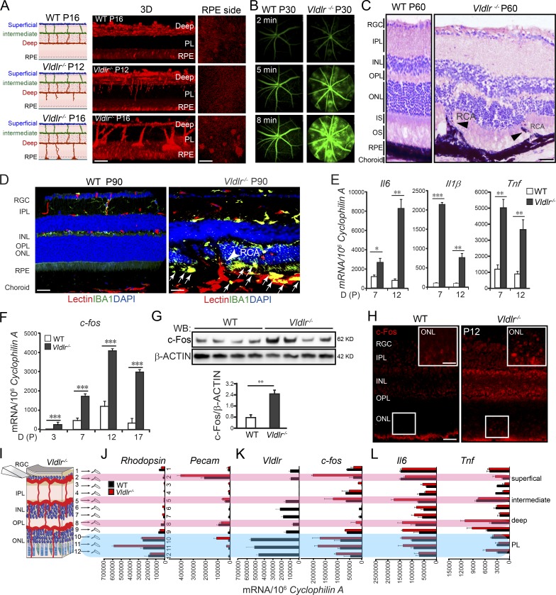
Figure 1.
c-Fos was induced in Vldlr−/− retinas. (A) Vldlr deficiency led to neovascularization in the normally avascular photoreceptor layer shown by 3D reconstruction of representative confocal images. n = 6. (B) Vldlr deficiency led to retinal vascular leakage at 2, 5, and 8 min after intraperitoneal injection of fluorescent dye, as shown by FFA images from P30 WT and Vldlr−/− mice. n = 6. (C) H&E staining showed retinal layer disorganization and retinal-choroidal vascular anastomoses in P60 Vldlr−/− retinas. Black arrowheads indicate neovascularization. n = 6. (D) Macrophage marker IBA1 (green) costained with the endothelial cell marker isolectin (red) and nuclear marker DAPI (blue) in 3-mo Vldlr−/− retinas with retinal-choroidal vascular anastomoses (arrowhead); macrophages were seen in the subretinal space between the ONL and RPE (arrows). (E) Cytokine expression, including Il6, Il1β, and Tnf, was increased during development in Vldlr−/− retinas. n = 6. (F–L) c-fos mRNA and protein expression were markedly increased (F and G) in Vldlr−/− retinas during development (F), mainly in the P12 ONL (K) and colocalized with the expression of Vldlr in P12 WT (H–K) and its target genes including Il6 and Tnf (L). n = 6. (H) IHC staining showed increased c-Fos expression in the ONL of P12 Vldlr−/− retinas. INL, inner nuclear layer; IPL, inner plexiform layer; IS, photoreceptor inner segment; OPL, outer plexiform layer; OS, photoreceptor outer segment; PL, photoreceptor layer; RCA, retinal-choroidal anastomosis; RGC, retinal ganglion cell; WB, Western blot. Bars: (A, 3D) 100 µm; (A, RPE) 250 µm; (C, D, and H) 50 µm; (H, inset) 25 µm. All data are representative of at least three independent experiments. *, P < 0.05; **, P < 0.01; ***, P < 0.001. Results are presented as mean ± SEM.

