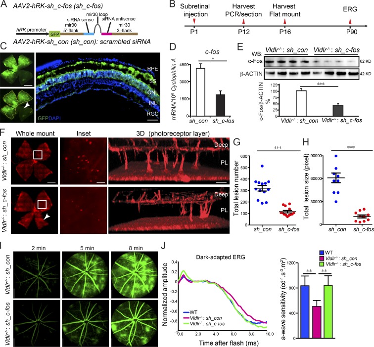
Figure 2.
Attenuation of c-Fos in the photoreceptor layer extending to the subretinal space reduced neovascularization. (A) Schematic diagram illustrating AAV2 carrying sh_c-fos within a mir30 cassette under the control of an hRK promoter (hRK-sh_c-fos). sh_con, sh_control. (B) Time line for AAV subretinal injection and retina collection. (C) The infection of AAV2 in the subretinal space (SS) and photoreceptor layer was confirmed by AAV2-hRK-eGFP. n = 6. INL, inner nuclear layer; RGC, retinal ganglion cell. The white arrowhead indicates the retinal areas were not affected by AAV2-hRK-eGFP because of the limitation of subretinal injection. (D and E) The knockdown efficiency of AAV2-hRK–sh_c-fos via subretinal injection into Vldlr−/− retinas was confirmed by comparison with AAV2-hRK-sh_control–injected retinas at both mRNA (D) and protein (E) levels. (D) *, P < 0.05. n = 6. (E) ***, P < 0.001. n = 4. WB, Western blot. (F) Knocking down c-fos in the photoreceptor layer (PL) extending to the subretinal space using AAV2-hRK–sh_c-fos–inhibited neovascularization shown by whole-mounted images of AAV2-hRK-sh_control– or AAV2-hRK–sh_c-fos–treated Vldlr−/− retinas stained with isolectin IB4 to label endothelial cells and representative 3D reconstruction images for neovascularization in photoreceptor layers, including the retinal areas, which may not be completely affected by AAV2-hRK–sh_c-fos (about 25% not transfected) as shown in C (white arrowhead in C and F). (G and H) Both total lesion number and total lesion size (pixels) were reduced in AAV-hRK–sh_c-fos–treated compared with AAV2-hRK-sh_control–treated retinas. ***, P < 0.001. n = 8–13. (I) AAV2-hRK–sh_c-fos injected in the subretinal space at P1 rescued Vldlr deficiency–induced retinal vascular leakage shown by FFA images at P60 in Vldlr−/−-treated mice. n = 6–8. (J) Representative dark-adapted (scotopic) ERG graph (left) and quantification of photoreceptor a-wave sensitivity (right) showed that photoreceptor function was attenuated in 3-mo-old Vldlr−/− retinas and was particularly rescued by AAV-hRK–sh_c-fos (one-time treatment on P1) in 3-mo-old Vldlr−/− retinas. *, P < 0.05; **, P < 0.01; ***, P < 0.001. Bars: (C and F, whole-mount images) 1,000 µm; (C, cross section) 50 µm; (F, inset) 250 µm; (F, 3D) 100 µm. All data are representative of at least three independent experiments. Results are presented as mean ± SEM.

