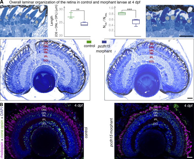
Figure 4.
Pcdh15 knockdown in X. tropicalis does not affect retinal morphogenesis. (A) Semithin sections of control and morphant retinas at 4 dpf. The retinal layer shows similar organization in morphant and control retina. However, a loss of alignment and shape alterations of the photoreceptor outer segments (OS) are seen in the morphants (see high magnification of the boxed areas). The outer retina is significantly thinner in the morphants: thickness of the OS, outer nuclear layer (ONL), and outer plexiform layer (OPL) (L(OS + ONL + OPL) = 24.6 ± 0.3 µm, mean ± SEM, in morphants versus 27.7 ± 0.6 µm in controls, n = 3–4; unpaired t test, **, P = 0.005. The ratio of the number of OS profiles (NOS) to that of nuclei (NONL) is also significantly lower in morphants (0.55 ± 0.05, mean ± SEM, in morphants versus 0.83 ± 0.02 in controls, n = 5; unpaired t test, ****, P = 0.0005). (B) Cryosections of 4 dpf retinas stained with antibodies against rhodopsin (magenta) and cone opsin (green) show no evidence of opsin mislocalization in the morphant retina, but the shape and organization of both cones and rods are altered. INL, inner nuclear layer; IPL, inner plexiform layer; GCL, ganglion cell layer. Bars, 20 µm.

