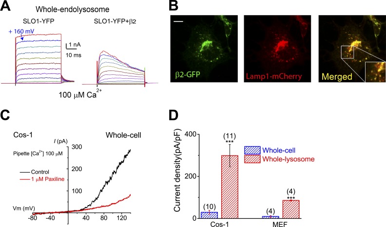
Figure 4.
LysoKVCa is preferentially expressed in the lysosome. (A) Step currents of Lyso-SLO1 (left) and Lyso-SLO1+ β2 (right) in the presence of 100 µM Ca2+. (B) Localization of β2-GFP in the Lamp1-positive compartments. Bar, 10 µm. (C) Whole-cell K+ currents sensitive to paxilline (1 µM) in nontransfected Cos-1 cells. The pipette solution contained 100 µM Ca2+. (D) Densities (mean ± SEM) of plasma membrane BK-like currents vs. LysoKVCa in Cos-1 cells and MEFs. Statistical comparisons were made with variance analysis (Student’s t test). ***, P < 0.001.

