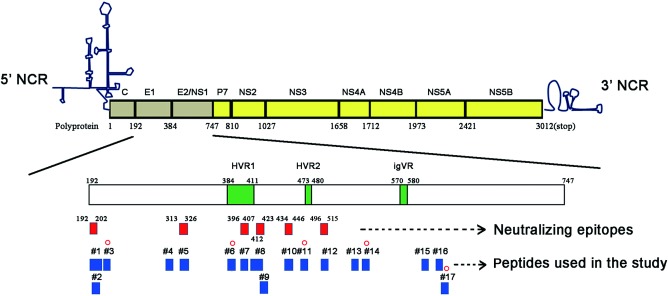
Figure 7.

Distribution of five defined linear natural killer‐mediated antibody‐dependent cellular cytotoxicity (NK‐ADCC) epitopes on hepatitis C virus (HCV)‐E1/E2. The position of HVR1 (aa384‐aa411), HVR2 (aa473‐aa480) and igVR (aa570‐aa580) in the open reading frame (ORF) of E1/E2 protein (aa192–aa747) are shown by green rectangles. Six putative neutralizing epitopes (aa192–aa202, aa313–aa326, aa396–407, aa412–aa423, aa434–aa446, and aa496–515) are indicated by red rectangles. The 17 linear epitope peptides used in this study are indicated by blue rectangles. NK‐ADCC‐specific epitopes (3, 6, 11, 14 and 17) identified in this study are marked with red circles. HVR = hypervariable region; igVR = intergenotypic variable region; NCR = non‐coding region. [Colour figure can be viewed at wileyonlinelibrary.com].
