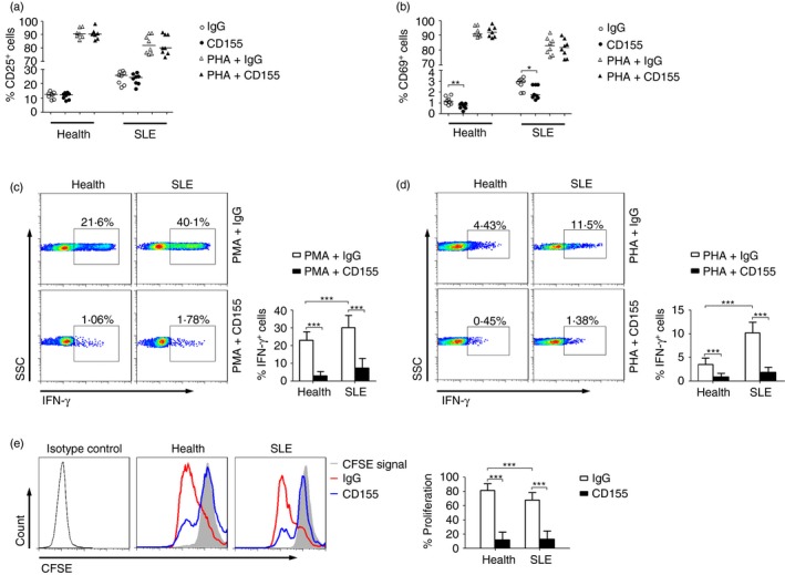
Figure 5.

Activation of TIGIT pathway down‐regulates the function of CD4+ T cells from systemic lupus erythematosus (SLE) patients. Peripheral blood mononuclear cells (PBMCs) isolated from healthy individuals and patients with SLE were stimulated with PMA or PHA in the presence of recombinant human CD155 protein or IgG control for 24 hr. (a) The percentages of CD25+ or (b) CD69+ cells in CD4+ T cells from healthy individuals (n = 7) and patients with SLE (n = 8) are shown. Each symbol represents an individual donor, and horizontal bars indicate the median. (c) Representative FACS plots showing the expression of interferon‐γ (IFN‐γ) in CD4+ T cells after PMA or (d) phytohaemagglutinin (PHA) stimulation. The percentages of IFN‐γ + cells in CD4+ T cells from healthy individuals and SLE patients are shown as the mean ± SD, n = 8 to n = 12 subjects per group, pooled from three independent experiments. (e) Representative FACS histograms showing the proliferation of CD4+ T cells from healthy individuals and SLE patients by using CD155 or IgG control. The percentages of CD4+ T‐cell proliferation in different groups are shown as the mean ± SD, n = 8 to n = 10 subjects per group, pooled from three independent experiments. *P < 0·05, **P < 0·01, ***P < 0·001 (Mann–Whitney U‐test). [Colour figure can be viewed at wileyonlinelibrary.com]
