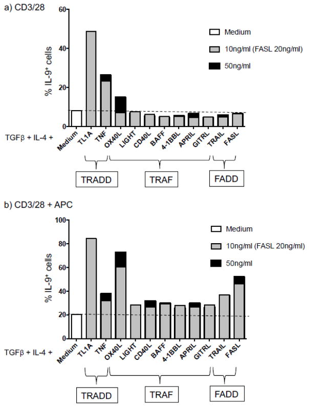
Fig. 2.
Mouse naïve CD4+ T cells were cultured in Th9-polarizing conditions (TGFβ + IL-4) in absence or presence of the indicated TNF family cytokines on coated plate with anti-CD3 and anti-CD28 in absence (a) or in presence of APCs (b) and were analyzed for their IL-9 expression by flow cytometry after restimulation with PMA and ionomycin for 4 hrs.

