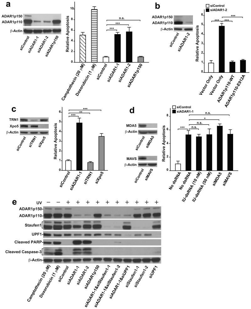
Figure 5. ADAR1p110 protects stressed cells from apoptosis.
(a-d) The extent of apoptosis was evaluated by ApoTox-Glo Triplex apoptosis assay (Promega), which simultaneously measures cell viability, cytotoxicity, and apoptosis (caspase3/7 activities). The signal intensity of relative luminescent units corresponding to caspase 3/7 activities was normalized with relative fluorescent units representing the cell viability measurements (400 nm/505 nm). More details in ONLINE METHODS. (a) ADAR1 knockdown induced apoptosis in UV-irradiated A172 cells at a much higher rate than in control cells. ADAR1p150 specific knockdown did not cause UV-induced apoptosis. For ADAR1 p150 selective knockdown, a specific siRNA targeting the human ADAR1 mRNA (NM_001111.4) exon1 region was used. Positive apoptosis controls: un-irradiated A172 cells treated with Camptothecin (20 μM) for 48 hrs or Doxorubicin (1 μM) for 24 hrs. (b) Apoptosis induced in UV irradiated cells by ADAR1 knockdown was rescued by transfection of both wildtype (WT) and editing deficient mutant (E912A) of ADAR1p110 expression vectors. For selective knockdown of endogenous ADAR1, siRNAs corresponding to the human ADAR1 mRNA 3′UTR (siADAR1-2) were designed and used here. (c) Apoptosis was induced in UV irradiated cells by Xpo5 knockdown but not by TRN1 knockdown. (d) Apoptosis induced in UV irradiated cells by ADAR1 knockdown was not rescued by transfection of synthetic I:U dsRNAs, MDA5 knockdown, or MAVS knockdown. (a-d) Knockdown efficiency by each siRNA was confirmed by immunoblotting (left panels). Data are mean ± S.D. (n = 4, cell cultures); significant differences were identified by two-tailed Student's t-tests: *P <0.05, **P < 0.01, ***P < 0.001, n.s., not significant. Source data are available online in Source Data Set 1. (e) The extent of apoptosis induced in UV-irradiated A172 cells was evaluated by western blotting analysis of cleaved PARP (89 kDa) and Caspase-3 (17 and 19 kDa) fragments. (a-e) Source data for western blotting images are available online in Source Data Set 2.

