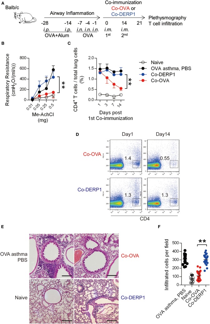
Figure 1.
Coimmunization alleviates airway inflammation through regulatory T cells. (A) Experimental scheme for coimmunization in the asthma model. Airway inflammation was induced from days −28 to 0 with OVA per marked dates. Mice were then coimmunized on day 0 and 14. Plethysmography and the infiltration were tested on day 21. Both antigen-matched immunization (Co-OVA) and mismatched immunization (Co-DERP1) were performed. (B) Plethysmography under methylcholine chloride stimulation. Naïve mice (black circle), PBS (black dot)-, Co-OVA (red dot)-, and Co-DERP1 (blue dot)-treated asthmatic mice are shown. n = 4. Same color labeling is used in similar settings there forth. (C) Cellular infiltration in lung tissues. Dynamics after coimmunization was analyzed with FACS. n = 4. (D) FACS data of T cell infiltration in the lung. (E) H&E histology of the lung infiltration on day 21. (F) Infiltrating cells per H&E slides were analyzed for each group. Every image represents an eye-field under microscope.

