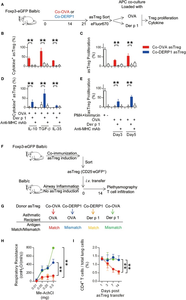
Figure 2.
Antigen-specific regulatory T cell (asTreg) transfer ameliorates antigen-matched asthma in vivo. (A–E) Coimmunization-induced asTregs show specificity in vitro. (A) Experimental scheme for in vitro re-stimulation of sorted asTregs. Foxp3-eGFP Tg mice were immunized on day −21 and −7. On day 0, asTregs were sorted by FACS (Figure S1B in Supplementary Material), labeled and cocultured with antigen-loaded APCs. Both antigen-matched (Co-OVA asTregs with OVA; Co-DERP1 with Der p 1) and mismatched ones (Co-OVA asTregs with Der p 1; Co-DERP1 with OVA) were performed. eFluor670 dilution was analyzed on day 3 and day 5. Cytokine secretion was analyzed 24 h post restimulation by FACS. (B,C) Co-OVA asTregs were re-stimulated with OVA or Der p 1. Intracellular suppressive cytokines secretion (b) and Treg proliferation (C) were analyzed. Anti-MHC-II mAb was added to block MHC-TCR signal. n = 4. (D,E) as above, Co-DERP1 asTregs were re-stimulated with OVA or Der p 1. n = 4. Data shown represent 3 independent experiments. (F) Experimental scheme for asthma treatment by asTreg transfer. (G) Antigen-matched and mismatched pairs of asTreg-transfer were performed. (H) Respiratory resistance of OVA asthmatic recipient mice (red and blue) or Der p 1 asthmatic recipients (orange and green) were analyzed on day 14 with plethysmography. n = 4. (I) Dynamics of CD4+ T cell presence in lung tissues by FACS. Data shown represent three independent experiments.

