Figure 7.
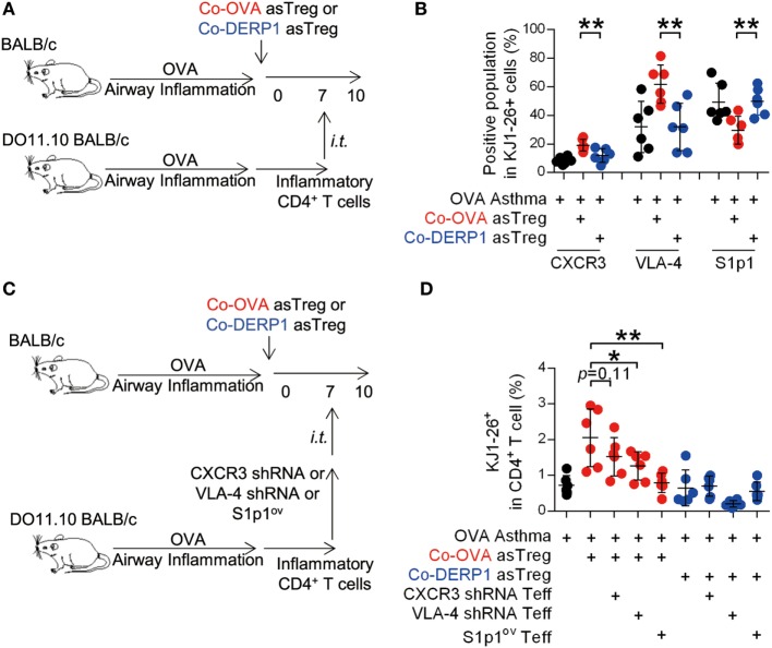
Antigen-specific regulatory T cells (asTregs) control the traffic of inflammatory effector T cells (Teffs) in hilar lymph node (hLN). (A) Similar to Figure 6A. 3 days after the i.t. transfer (day 10), chemokine receptors and adhesion molecules were analyzed on KJ1-26+ cells in hLN. Both antigen-matched (Co-OVA, red) and mismatched (Co-DERP1, blue) treatments were analyzed. (B) Enrichment of the CXCR3+, VLA-4+, S1p1− population in DO11.10 T cells in hLN. n = 6. (C) Experimental scheme for lentivirus knockdown assay. CXCR3 and VLA-4 knockdown, and S1p1-overexpression were performed on donor inflammatory DO11.10 T cells, and they were i.t. transferred into asthmatic asTreg-recipient mice on day 7. (D) On day 10, KJ1-26+ cells recruited back to hLN were analyzed. n = 6. Data shown represent two independent experiments.

