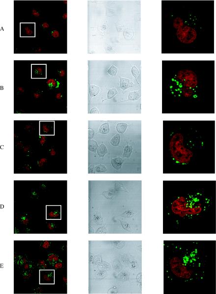
Figure 4.
Confocal microscope images of the free uptake of fluorescein-labelled CPP conjugates (green) into HeLa cells. Left panels show a range of cells (nucleus stained red). Central panels are the same cells in DIC. Right panels show a magnification of a single cell boxed in the left panels. Horizontally, (A) conjugate 4 (OMe/LNA oligo conjugated with the C-terminus of Tat peptide), (B) conjugate 6 (OMe/LNA oligo B conjugated with the C-terminus of Penetratin), (C) conjugate 5 (OMe/LNA oligo B conjugated with the N-terminus of Penetratin), (D) conjugate 12 (OMe/LNA oligo B conjugated with R9F2) and (E) conjugate 10 (PS/OMe oligo C conjugated with the C-terminus of Penetratin).

