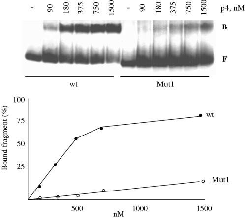
Figure 7.
Binding of p4 to the Mut1 sequence. Increasing amounts of p4 were incubated in 20 μl of binding buffer with a fixed amount of fragment containing the 31 bp of the inverted repeat, corresponding to site 1 or Mut1 sequence surrounded by 31 bp of identical but unspecific double-stranded DNA. B denotes the protein–DNA complexes and F, free DNA. Below, representation of the percentage of p4–DNA complexes formed by increasing concentration of p4 with the wild-type (wt) or mutated site 1 (Mut1).

