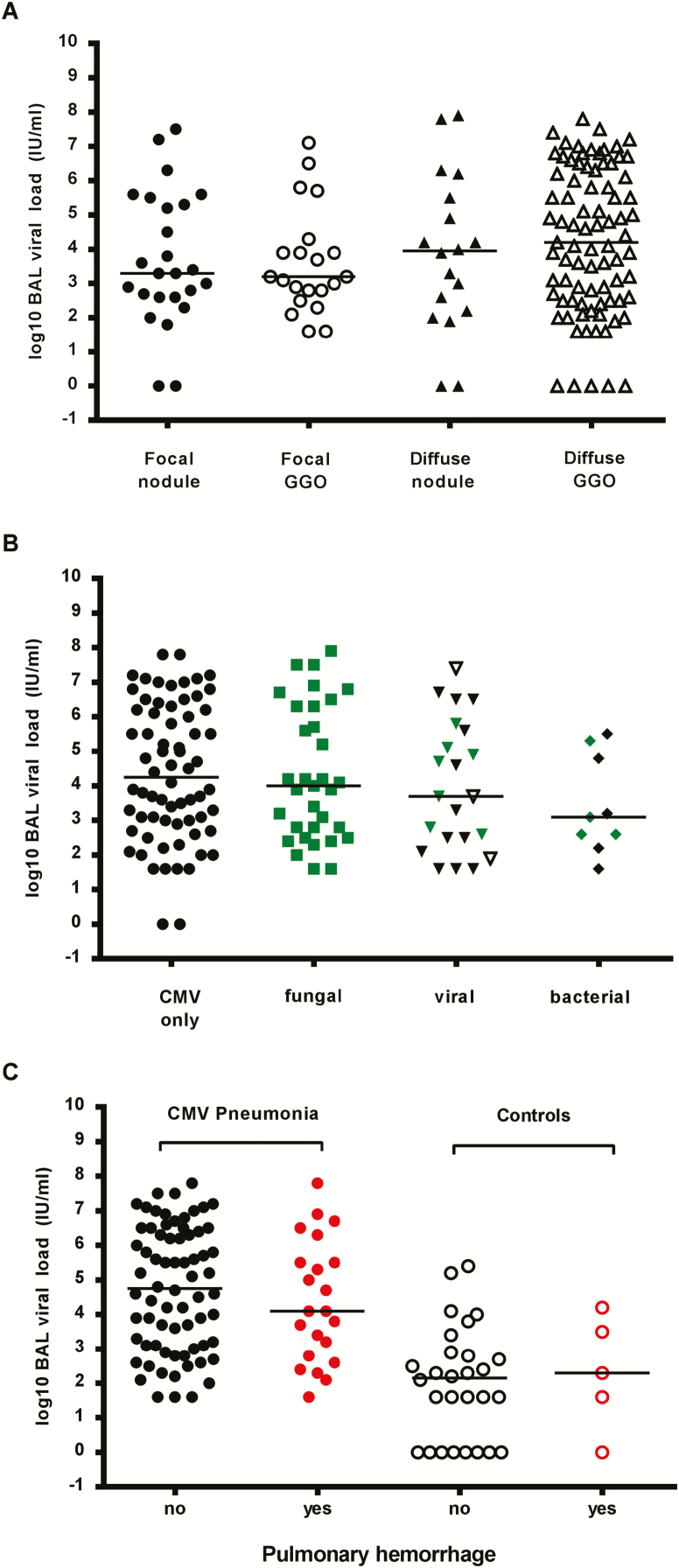
Figure 2.
Quantitative cytomegalovirus (CMV) load in bronchoalveolar lavage (BAL) fluid according to possible effect modifiers. A, Viral load in BAL fluid from CMV pneumonia cases did not differ with respect to the radiologic appearance of the lungs at the time of the BAL. Radiographic presentation was categorized by a pulmonologist blinded to the CMV results in the following categories: (1) focal nodule(s), (2) focal ground-glass opacities (GGOs), (3) diffuse nodules, (4) diffuse GGOs. Outcomes in subjects with non-CMV pneumonia with a high viral load were evaluated by chart review. If multiple locations were lavaged on the same day, each is represented separately. One CMV pneumonia case with multiple BALs on the same day was excluded because BAL polymerase chain reaction (PCR) results could not be assigned to a specific BAL location. A total of 147 BALs were performed on 131 patients. B, Quantitative CMV load in BAL fluid from CMV pneumonia cases did not differ according to the absence (CMV only) or presence of any specific copathogen subset. Detection of Aspergillus galactomannan in the BAL or blood counted as detection of a fungal copathogen. Co-occurrence of fungal copathogens with other copathogens is denoted by green markers. If bacteria were present in addition to a viral copathogen, the BAL finding is grouped with viral copathogens but is denoted with an open marker. A total of 68 subjects had CMV only detected, 33 had a fungal copathogen detected, 22 had a viral copathogen detected, and 9 had a bacterial copathogen detected. C, Viral loads in cases and controls were similar in the presence or absence of pulmonary hemorrhage in patients with CMV DNAemia within 7 days of BAL (data are for 92 patients with CMV pneumonia and 33 controls).

