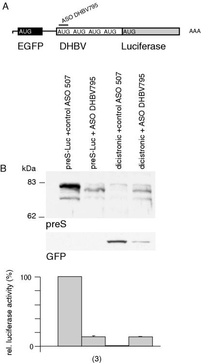
Figure 6.
The DHBV preS region does not display IRES activity. (A) Schematic diagram of a dicistronic vector comprising EGFP and preS Luc is shown. (B) Immunoblotting of preS luciferase fusion proteins and EGFP upon cotransfection with ASO. Monocistronic preS-Luc served as positive control. Equal amounts of protein were loaded. Relative luciferase activity was compared to luciferase activity obtained after cotransfection of the monocistronic construct with control ASO and is given in percent. The number of observations is given in brackets.

