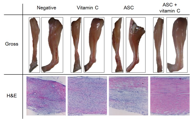
Figure 3. Gross and microscopic features of tendons. Gross view: The vitamin C depletion groups exhibited more neovascularization, swelling, and mucus secretion than the groups supplied with vitamin C. Histopathology: The control group exhibited worsened lesions, indicated by separation of fibers with loss of bundle demarcation, high cell density, tenocytes with round nuclei, and infiltration of inflammatory cells. Tenocytes with round nuclei and immature collagen fibers were observed in the group treated with vitamin C, but demarcated bundles were maintained. The group treated with adipose-derived stem cells (ASCs) exhibited local tendon healing (upper part). The combination group exhibited nearly normal tendon features, such as well-demarcated collagen bundles, elongated tenocytes, mature collagen fibers, and low cell density. Gross view: Front and lateral views; histopathology: Hematoxylin and eosin staining, magnification ×100.

