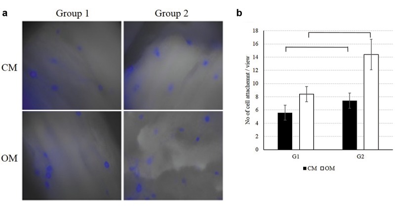
Figure 2. Cell attachment on the surface of duck-beak bone. Images of cell attachment were taken by a fluorescence microscope (a). Group 2 has a significantly higher number of attached cells compared with Group 1 (b) (×400). CM, Control media; OM, osteogenic media.

