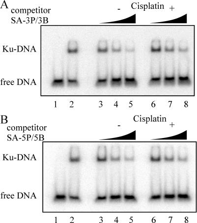
Figure 3.
Effect of strand bias, sequence specificity and cisplatin–DNA adduct position on Ku binding. (A) Competition binding assays were performed in 20 μl reactions and contained 2.5 nM 32P-labeled double-stranded 2.1/2.2 DNA and 5 nM Ku. Increasing concentrations of the SA-bound competitor DNA (3P/3B), 5, 50 and 200 nM is indicated by the triangles. Controls for the assay include 32P-labeled double-stranded 2.1/2.2 DNA alone (lane 1) and 32P-labeled double-stranded 2.1/2.2 DNA with 5 nM Ku in the absence of any competitor (lane 2). (B) Competition assays were carried out identically to those in (A) except reactions included 5P/5B competitor DNA.

