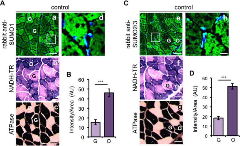
Fig. 2.
Immunofluorescence analysis of SUMO conjugates in control diaphragm. A and C, muscle fibers are differently stained with antibodies to SUMO1 and SUMO2/3. Cellular localization of free and conjugated SUMO1 (A, panel a) and SUMO2/3 proteins (C, panel e) on cryo-cross-sections were detected by immunofluorescence with anti-SUMO1 and anti-SUMO2/3 antibodies raised in rabbit and with secondary anti-rabbit AlexaFluor 488-conjugated antibodies. The white box areas are enlarged (A, panel d, and C, panel h), showing the nuclear localization of both SUMO1 and SUMO2/3. A, panel b, and C, panel f, consecutive serial muscle sections were subjected to NADH-TR staining to recognize the oxidative (O, dark purple) and glycolytic (G, light purple) fibers. A, panel c, and C, panel g, representation of ATPase, pH 10.3, staining to distinguish type I (white) and type II (black) fibers. B and D, mean fluorescence intensity per area and ±S.D. for SUMO1 (B) and SUMO2/3 (D) immunofluorescences were measured considering a number of 35 oxidative and 35 glycolytic fibers, respectively, from four different diaphragm control sections (***, p < 0.001) and expressed in AU. Scale bars, 50 μm (panels a–c and panels e–g) and 20 μm (panels d and h). Nuclei are stained with DAPI (blue). TR, Tetrazolium Reductase.

