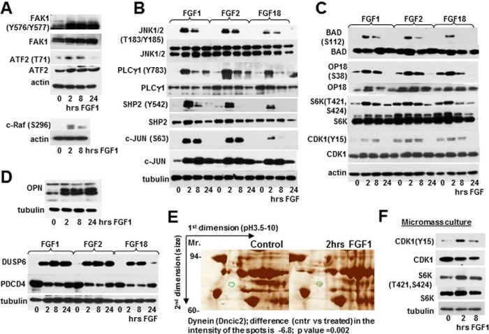
Fig. 4.
Validation of FGF regulated targets. RCS cells (A–E) and micromass cultures (F) were treated with FGF1, FGF2, and FGF18 as marked, and analyzed by immunoblotting using indicated antibodies. Twenty micrograms of total protein from RCS lysates and 10 μg of total protein from micromass lysates was used for immunodetection. The blots are representative of at least three independent experiments. E, Fragments of silver stained 2D electrophoresis. RCS cells were treated with FGF1 for 2 h and lysates obtained from treated and untreated cells were enriched for phosphoproteins following 2D electrophoresis. An identified protein those phosphorylation status is changed upon FGF1 treatment is circled in green. Two independent experiments were performed with similar results.

