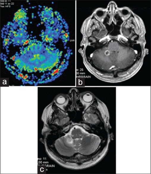
Figure 5.

(a) M2: MR Perfusion imaging at 8.5 months (M2): No increase in CBV. (b) M2: Slight increase in tumor volume / central necrosis on MRI CE T1. (c) M2: Limited perilesional edema on MRI T2 with intermittent cortisone treatment: probable focal ARE
