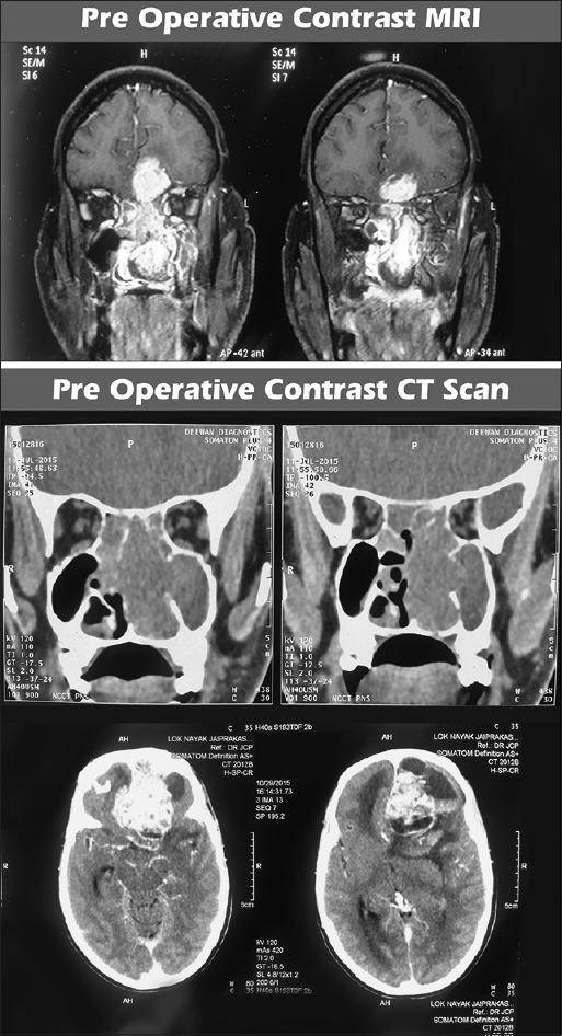
Figure 1.

Pre operative CEMRI and CECT scan of the brain and paranasal sinuses showing dumbbell shaped mass in the left superior nasal cavity with extensions into the anterior cranial fossa and paranasal sinuses. The ‘waist’ of the dumbbell is at the cribriform plate. The mass shows intense heterogeneous post contrast enhancement. Peritumoral cysts are also noted at the tumor brain interface
