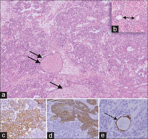
Figure 3.

(a and b) Hematoxylin and eosin stained slides (x100) - showing olfactory neuroblastoma which is having a lobular pattern of primitive cells, no fibrillary matrix and showing squamous (single arrow) and glandular differentiation (double arrow) and inset (x200) having true rosettes (Flexner Wintersteiner rossette, double head arrow). (c-e) IHC (×400) (c) Strong synaptophysin positivity is seen in the tumor cells, (d) Cytokeratin 5/6 positivity is seen in the squamous cells and (e) Cytokeratin 7- focal positivity in the glands (thin arrow)
