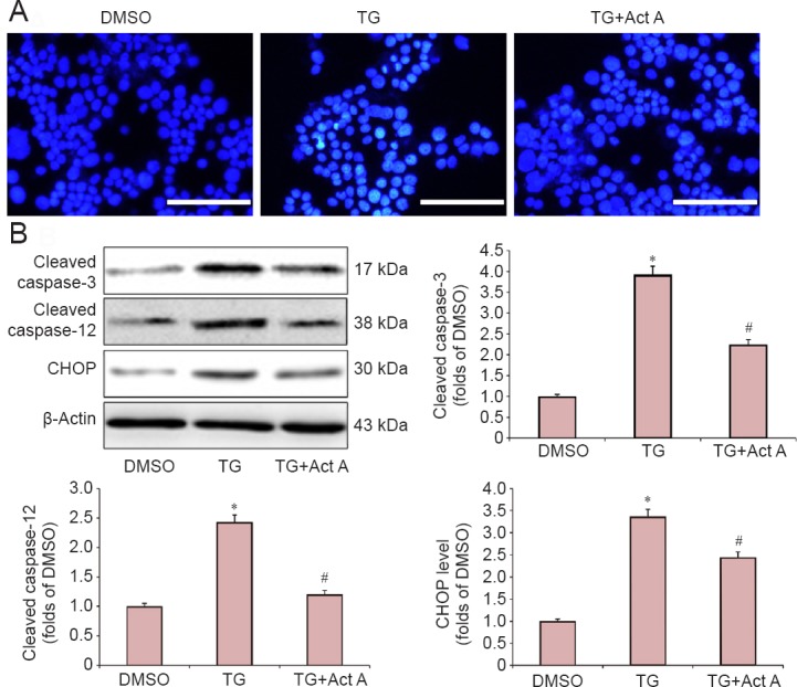
Figure 2.

Act A reduced endoplasmic reticulum stress-induced apoptosis in PC12 cells.
Cells were pretreated with Act A (100 ng/mL) for 24 hours, and then co-treated with TG for 20 hours. (A) The morphological features of apoptosis were monitored by fluorescence microscopy after staining with Hoechst 33342. Scale bars: 100 μm. (B) Western blot assay of the expression of cleaved-caspase-3, cleaved-caspase-12 and CHOP. Protein expression was quantified by optical density and normalized to β-actin. The fold change compared with the DMSO group for each protein is expressed as the mean ± SD of three independent experiments. One-way analysis of variance followed by Bonferroni post hoc tests were used for statistical analysis. *P < 0.05, vs. DMSO group; #P < 0.05, vs. TG group. DMSO: Dimethyl sulfoxide; TG: thapsigargin; Act A: Activin A.
