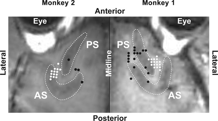
Fig. 3.
Structural MR images tangential to the surface of the cortex underlying the recording chambers. White dots indicate recording sites at which task-related neurons were encountered. Black dots indicate recording sites at which no task-related neurons were encountered. Images from the 2 monkeys abut at the interhemispheric midline. AS, arcuate sulcus. PS, principal sulcus.

