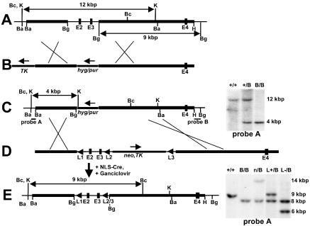
Figure 1.
Targeted mutagenesis at the Rev1 BRCT domain. (A) The 5′ region of the genomic Rev1 locus containing exons 2, 3 and 4 (E2–E4). Horizontal bold sections, genomic regions homologous to the targeting vector. (B) Targeting vectors pBRCT–hygro and pBRCT–puro to delete the Rev1 gene exons 2 and 3 encoding most of the BRCT domain. Hyg, pPGK–hyg cassette. Pur, pPGK–pur cassette. TK, pPGK–thymidine kinase cassette. Arrows, direction of transcription. (C) Targeted Rev1B (hyg or pur) alleles. Probes A and B, DNA fragments used for analysis of gene targeting events. At the right side of the map, a Southern blot of genomic DNA of targeted ES cell lines, digested with KpnI, is depicted. +/+: wild-type ES cells. +/B: Rev1+/B (hyg) cells. B/B: Rev1B/B (hyg, pur) ES cells. (D) pREV1–loxP targeting vector used for constructing the marker-less Rev1L− allele and for restoration of the Rev1B (hyg) allele to wild type. L1: upstream loxP site. Neo, TK: pPGK–neomycin and pPGK–thymidine kinase cassettes, flanked by loxP sites (L2 and L3). (E) Rev1 genomic locus of the Rev1L+/B (pur) ES cell line. At the right side of the map, a Southern blot of genomic DNA of targeted ES cell lines, digested with BclI, is shown. Sizes of fragments representing the different Rev1 alleles are the following: 14 kb: Rev1B (neo/TK), 9 kb wild type, 8 kb Rev1B (pur), and 6 kb Rev1L-. +/+: wild-type ES cells. B/B: Rev1B/B ES cells. n/B: Rev1n/B (neo/TK, pur) ES cells.L+/B: Rev1L+/B (pur) ES cells. L−/B: Rev1L−/B (pur) ES cells. Ba, BamHI; Bc: BclI; Bg, BglII; H, HindIII; K, KpnI.

