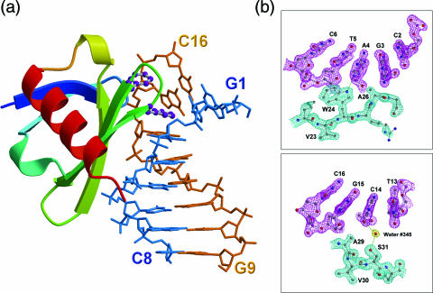
Figure 1.
Sac7d-DNA interface. (a) Ribbon diagram of Sac7d V26F/M29F-GCGATCGC complex with two intercalating phenylalanine residues depicted as ball and stick. The aromatic ring of Phe26 residue stacks with the G3 base, whereas the phenyl ring of Phe29 is stacked on the deoxyribose of G15. (b) The (2Fo − Fc) Fourier electron density maps of the V26AM29A–GCGATCGC complex (contoured at 1.5σ level) in the regions at the protein–DNA interface (upper panel). The indole NH group of Trp24 forms hydrogen bond (3.0 Å) to the base N3 of A4. Adjacent to the intercalating site, Ser 31 (OG) forms a water-mediated hydrogen bond with C14 O2 (lower panel).

