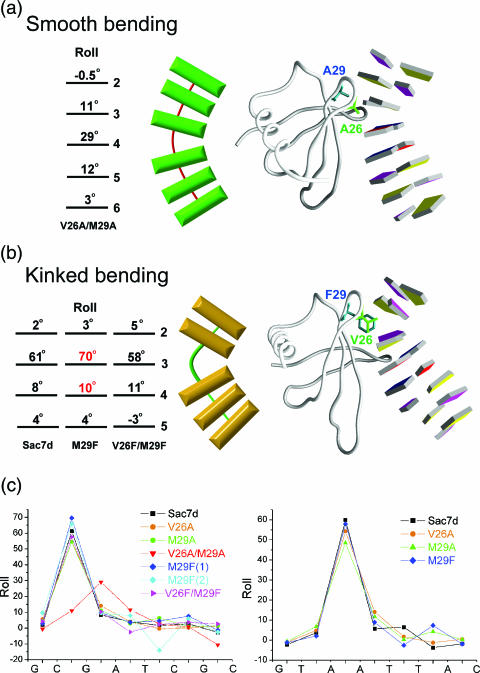
Figure 2.
Two modes of DNA bending. (a) The structure of V26A/M29A–DNA complex (right) and schematic diagram (left) stress gradual curvature by smooth bending. (b) The structure of M29F–DNA complex shows localization of curvature by kinked bending from minor groove of DNA. The contributions of each base-pair-step curvature to overall helix bending are listed with corresponding base-pair numbers. (c) Local base-pair step parameter, roll, for all eight Sac7d mutant–DNA binary structures, calculated using the X3DNA program. Note the largest roll at certain kinked step (G3pA4 in V26A/M29A, C2pG3 or A3pA4 in the others).

