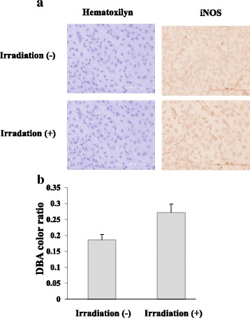
Fig. 6.

Expression of iNOS in Ca9–22 cells by irradiation with the 310 nm UVB-LED. Monolayers of Ca9–22 cells in a chamber slide were irradiated by the 310 nm UVB-LED for 60 s and incubated for 24 h. a Expression of iNOS in the cells was measured by immunofluorescent staining using an anti-iNOS antibody. b The levels of DAB color ratio were measured by using Image J. (n = 3, means ± SE; P = 0.0503)
