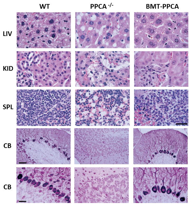
Figure 4. Histology of systemic organs and cerebellum from BM-transplanted GS mice.

Top panels: Organs from PPCA −/− mice transplanted with total −/− BM transduced with the MSCV-PPCA (BMT-PPCA) retrovirus were isolated at different time points after treatment. Hematoxylin and eosin-stained tissue sections of the liver (LIV), kidney (KID), and spleen (SP) from a BMT-PPCA–treated PPCA −/− mouse sacrificed 9 months after treatment, and from age-matched wild-type and PPCA −/− mice revealed the complete restoration of normal tissue morphology with BM expressing PPCA, compared to the extensive vacuolation present in the PPCA −/− control mouse. Size bar corresponds to 30 μm. Lower panels: Serial sections of the cerebellum from a 9-month-old GS mouse transplanted with MSCV-PPCA–marked BM cells were stained with anti-PEP19 antibody. Note the dramatic loss of Purkinje cells in an age-matched GS mouse and the significant number of these cells that are retained in the treated animal. Size bars correspond to 60 μm and 30 μm Adapted from Leimig et al 2002 Blood, 58 with permission of the American Society of Hematology.
