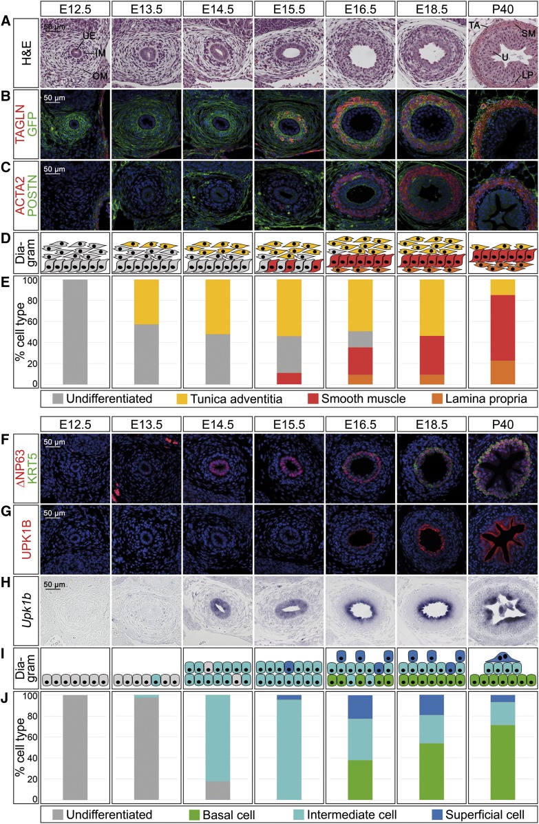
Figure 1.
Cell differentiation in ureter development occurs in a temporally and spatially controlled manner. (A–E) Time course of cell differentiation in the ureteric mesenchyme. (A) Hematoxylin and eosin (H&E) staining on transverse sections of the proximal ureter. At E12.5, the inner and outer mesenchymal region (IM and OM) and the ureteric epithelium (UE) are indicated. At P40, the three subregions of the mesenchyme, the lamina propria (LP), the smooth muscle (SM), and the tunica adventitia (TA) are marked adjacent to the urothelium (U). (B and C) Coimmunofluorescence analysis of expression of the SMC marker TAGLN and the lineage marker GFP (B) and of the SMC marker ACTA2 and the adventitial marker POSTN (C) on transverse sections of the proximal ureter in Tbx18cre/+;R26mTmG/+ mice. Nuclei are counterstained with DAPI (blue). (D) Schematic representation of cell differentiation in the ureteric mesenchyme. Fibrocytes of the tunica adventitia (yellow) are defined as POSTN+GFP+, SMCs (red) as TAGLN+ACTA2+GFP+, and lamina propria cells (orange) as TAGLN−GFP+. (E) Quantification of differentiated cell types in the ureteric mesenchyme on the basis of marker expression as explained in (D). For numbers see Supplemental Table 1A. (F–J) Time course of epithelial differentiation in the ureter. (F and G) Coimmunofluorescence analysis of expression of the B cell marker KRT5, the B and I cell marker ∆NP63 (F), and the I and S cell marker UPK1B (G) with DAPI-stained nuclei (blue), and (H) in situ hybridization for Upk1b expression on transverse sections of the proximal ureter in Tbx18cre/+;R26mTmG/+ mice. (I) Schematic representation of cell expansion and differentiation in the ureteric epithelium. B cells (green) are defined as KRT5+∆NP63+UPK1B−Upk1b−, I cells (turquois) as KRT5−∆NP63+UPK1B+(low)Upk1b+, and S cells (blue) as KRT5−∆NP63−UPK1B+(high)Upk1b+. (J) Quantification of differentiated cell types in the ureteric epithelium on the basis of marker expression as explained in (I). For numbers see Supplemental Table 1B.

