Figure 4.
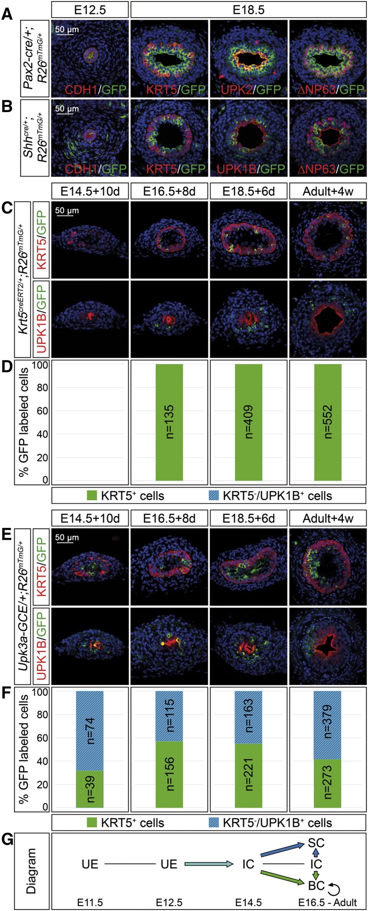
I cells are progenitors of B and S cells in ureter development and homeostasis. (A and B) Coimmunofluorescence analysis on transverse sections of the proximal ureter of the lineage marker GFP with the epithelial marker CDH1 at E12.5, and the B cell marker KRT5, the B and I cell marker ∆NP63, and the S cell markers UPK1B/UPK2 in Pax2-cre/+;R26mTmG/+ embryos (A) and in Shhcre/+;R26mTmG/+ embryos (B) shows that Pax2+ and Shh+ cells of the early ureteric bud contribute to all urothelial cell types in the ureter but not the surrounding mesenchyme. (C and E) Coimmunofluorescence analysis of the lineage marker GFP with the B cell marker KRT5 (upper panel) and the S cell marker UPK1B (lower panel) on transverse sections of ureters isolated at E14.5, E16.5, and E18.5, induced with 500 nM 4-hydroxytamoxifen for the first 24 hours and cultured for 10, 8, and 6 days; and in adult ureters 4 weeks after tamoxifen administration in Krt5creERT2/+;R26mTmG/+ mice (C) and Upk3a-GCE/+;R26mTmG/+ mice (E). (D and F) The bar diagrams display the percentage of lineage-labeled (GFP+) cells contributing to KRT5+UPK1B− B cells or to KRT5−UPK1B+ I and S cells in the ureter of Krt5creERT2/+;R26mTmG/+ mice (D) and of Upk3a-GCE/+;R26mTmG/+ mice (F) at the indicated stages and conditions. The number of counted GFP+ cells (n) is given. For additional numbers see Supplemental Table 4, A and B. (G) Schematic representation of the lineage relations of urothelial cell types. KRT5+ cells only give rise to B cells whereas UPK3A+ cells give rise to B, I, and S cells. BC, basal cell; IC, intermediate cell; SC, superficial cell; UE, undifferentiated epithelium.

