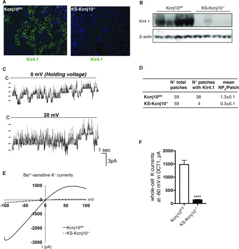
Figure 2.
K+ channel activity is reduced in KS-kcnj10−/− mice. (A) Representative immunostaining of Kir4.1 in kcnj10fl/fl and KS-kcnj10−/− mice. Original magnification, ×100. (B) Western analysis of Kir4.1 from kcnj10fl/fl and KS-kcnj10−/− mouse kidneys. Actin was the loading control. (C) Single channel recording of a 40 pS inwardly-rectifying K channel from the basolateral membrane of DCT1 in control mice. The experiments were performed in a cell-attached patch with 140 mM K in the pipette and 140 mM Na/5 mM K in the bath. The closed level is indicated by a dotted line and “C.” (D) The probability of finding the 40 pS K channel and channel activity (NPo) in DCT1 of kcnj10fl/fl and KS-kcnj10−/− mice. (E) Whole-cell recording showing Ba2+-sensitive K+ currents in DCT1 of kcnj10fl/fl (solid line) and KS-kcnj10−/− KO (dotted line) mice. The experiments were performed with a ramp protocol from −100 to 100 mV with symmetrical 140 mM KCl in the bath and in the pipette. (F) Bar graph summarizing the above results measured at −60 mV (n=5) is shown in the bottom panel. ***P<0.001 by t test.

